CELL BIOLOGY
PREPARED BY MR.
ABHIJIT DAS
CELL: cell is the
fundamental structural and functional unit of all living organisms. A cell
consists of a plasma membrane enclosing a number of organelles suspended in a
watery fluid known as cytosol.
PROTOPLASM:
The living part of a cell surrounded by a plasma membrane. (all inside plasma
membrane)
CYTOPLASM:
The semifluid substance of a cell excluding the nucleus. (protoplasm – nucleus)
The human body contains about 100 trillion cells.
HISTORY
Robert Hooke discovered
dead plant cell in 1665. However, it was Robert
Hooke who coined the term cell.
Antonie Van Leeuwenhoek discovered
living cell in 1674.
Robert brown discovered
nucleus of the cell in 1831.
CELL MEMBRANE
Cell membrane or the plasma membrane is the
protective semipermeable membrane, covering the cell body.
The cell membrane is composed of lipids that are arranged in a bilayer.
The lipids are arranged within the membrane with the
polar head (hydrophilic head) towards the outer
sides and the non-polar tails (hydrophobic
tails) towards the inner side.
This ensures that the hydrophobic tail is protected
from the aqueous environment.
The cell membranes also possess protein and carbohydrate.
Membrane proteins can be classified as integral or peripheral proteins.
Peripheral proteins lie on the surface of membrane
while the integral proteins are partially or totally buried in the membrane.
According to FLUID MOSAIC MODEL, the fluid nature of
lipid enables movement of proteins within the bilayer.

The most important function of plasma membrane is
the transport of the molecules across it.
DIFFERENT ORGANELLES OF CYTOPLASM:
1. Endoplasmic
reticulum
2. Golgi
bodies
3. Ribosome
4. Mitochondria
5. Lysosome
6. Centrioles

STRUCTURE AND FUNCTION OF CYTOPLASMIC
ORGANELLES
1. ENDOPLASMIC
RETICULUM
STRUCTURE
Ø Endoplasmic
reticulum is a network of tiny tubular structures scattered in the cytoplasm.
Ø They
are of two types: smooth and rough.
Ø The ER bearing ribosomes on their surface is called rough endoplasmic reticulum (RER).In the absence of ribosomes they appear smooth and are called smooth endoplasmic reticulum (SER).

FUNCTION
Ø RER
is involved in protein synthesis.
Ø SER
is the major site for synthesis of lipid and lipid-like
steroidal hormones.
2. GOLGI
APPARATUS
STRUCTURE

They consists of many flat, disc-shaped sacs which are called as cisternae.
FUNCTION
Ø A
number of proteins synthesized by ribosomes on the endoplasmic reticulum are modified (or processed) in the golgi apparatus.
Ø Golgi
apparatus is also the important site of formation of glycoproteins and
glycolipids.
3. MITOCHONDRIA
STRUCTURE

Ø Typically
mitochondria is cylindrical shaped having a
diameter of 0.2 - 1.0 micrometer and length 1.0 - 4.1micrometer.
Ø Mitochondria
is a double membrane bound structure and is described as the power house of the cell.
Ø The
inner compartment is called the matrix.
Ø The
inner membrane forms a number of foldings called as cristae
(singular: crista). It increases the surface area.
FUNCTION
Ø They
are the sites of aerobic respiration.
Ø They
produce cellular energy in the form of ATP hence they are called ‘power houses’
of the cell.
4. RIBOSOMES
They are tiny granules composed of
RNA and protein.
FUNCTION
They synthesize proteins from amino
acids.
5. LYSOSOMES
They are membrane bound vesicular
structures pinched off from the golgi apparatus.
FUNCTIONS
Ø They
are rich in almost all types of hydrolytic enzymes (lipases, proteases,
carbohydrases). These enzymes are capable of digesting macromolecules such as
carbohydrates, proteins, lipids etc.
Ø They
also break down fragments of organelles inside the cell into small particles
that are either recycled or removed as waste material.
Ø Lysosomes
in white blood cell (WBC) contain enzymes that digest microbes.
6. CENTROSOME
Centrosome is an organelle usually
containing two cylindrical structures called centrioles. Both centrioles lie
perpendicular to each other.
FUNCTION
They form spindle
fibres during cell division in animal cells.
NUCLEUS
Nucleus is present in almost all eukaryotic cells.
The nucleus is the largest
organelle and is covered by the nuclear membrane,
a double-layered membrane with tiny pores through which some substances can pass between
nucleus and the cytoplasm.
The outer layer of nuclear membrane is continuous
with endoplasmic reticulum.

The nucleus contains the body’s genetic material in
the form of DNA.
In a non-dividing cell,
DNA is present as a fine network of threads called chromatin,
but when the cell prepares to divide, the
chromatin forms compact structures called chromosomes.
RNA is also found in the nucleus which is involved
in protein synthesis.
Within the nucleus there is another spherical
structure called the nucleolus, which is
responsible for synthesis of ribosomes.
CELL CYCLE
The sequence of events by which a cell duplicates
its genome, synthesizes the other constituents of the cell and eventually
divides into two daughter cells is termed as cell cycle.
Cell growth results in disturbing the ratio between
the nucleus and the cytoplasm. That’s why it becomes essential for the cell to
divide to restore the nucleo-cytoplasmic ratio.
PHASES OF CELL CYCLE
The cell cycle is divided into two basic phases.
·
Interphase (95%
of the duration of the cell cycle)
·
M-Phase (5%
of the duration of the cell cycle)
INTERPHASE
The interphase represents the phase where the cell
prepares itself. That’s why it is otherwise called as preparatory
phase.
The interphase is divided into three further phases.
·
G1 Phase
·
S Phase
·
G2 Phase
During G1 Phase,
the cell continuously grows (cell growth).
During S Phase, DNA replication takes place. During this time the
amount of DNA per cell doubles. If the initial amount of DNA is denoted as 2C then
it increases to 4C, but there is no increase in the chromosome number.
During G2 Phase, proteins are synthesized in preparation for
mitosis while cell growth continues.
M-PHASE
The M-Phase represents the phase when the actual
cell division occurs. The M-Phase starts with the nuclear division (karyokinesis) and usually ends with division of
cytoplasm (cytokinesis). It is also called
as equational division.
In humans, mitotic cell division is only seen in the
diploid somatic cells.
KARYOKINESIS
Karyokinesis is divided into the following four
stages
·
Prophase
·
Metaphase
·
Anaphase
·
Telophase
PROPHASE
Ø Chromosomal material condenses. Chromosomes
are seen to be composed of two chromatids attached together at the centromere.


At
the end of prophase cells don’t show golgi
complexes, endoplasmic reticulum, nuclear membrane etc.

METAPHASE
Ø Spindle
fibres attach to kinetochores of
chromosomes.

Ø Chromosomes
are moved and get aligned along metaphase plate through
spindle fibres.

ANAPHASE
Ø Centromeres split and chromatids
separate.
Ø Chromatids
move to opposite poles.

TELOPHASE
Ø Chromosomes gather at opposite poles.
Ø Nuclear membrane reappears around the chromosomes.
Ø Other cellular organelles reappear.

CYTOKINESIS
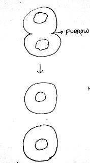
The cell is divided into two daughter cells by a
process known as cytokinesis at the end of which cell division is complete.
This is achieved by the appearance of a furrow in the plasma membrane. The furrow gradually deepens and ultimately joins in the centre dividing the cell into two.
SIGNIFICANCE OF MITOSIS
Ø The
growth of humans is due to mitosis.
Ø Another
significant contribution of mitosis is cell repair.
The cells of upper layer of epidermis, cells of the lining of the GIT, blood
cells etc. are being constantly replaced.
MEIOSIS
The production of offspring by sexual reproduction
includes the fusion of two gametes (sperm, eggs),
each with a complete haploid set of chromosomes.
Gametes are formed from specialized diploid cells (spermatogonia,
oogonia).
This specialised kind of cell division reduces the
chromosome number by half results in production of haploid daughter cells. This
kind of division is called meiosis.
Meiosis involves two sequential cycles of
karyokinesis and cytokinesis called meiosis I and meiosis II but only a single
cycle of DNA replication.
Four haploid cells are formed at the end of meiosis
II.
MEIOSIS I
PROPHASE I
It has been further subdivided into five phases
I.
Leptotene
II.
Zygotene
III.
Pachytene
IV.
Diplotene
V.
Diakinesis
LEPTOTENE

The condensation of
chromosomes continues throughout leptotene.
ZYGOTENE

During this stage the homologous chromosomes start
pairing together and this process is called synapsis.
The complex formed by a pair of synapsed homologous
chromosomes is called a bivalent or tetrad.
There is also a formation of complex structure
called synaptonemal complex.
PACHYTENE

At this stage crossing over occurs between
non-sister chromatids of the homologous chromosomes.
Crossing over leads to recombination of genetic
materials of the two chromosomes.
DIPLOTENE
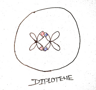
During this stage, the dissolution of synaptonemal
structure occurs.
The recombined homologous chromosomes separate from
each other except at the sites of crossovers.
These X shaped structures are called chiasmata.
DIAKINESIS

During this state, terminalisation of chiasmata
occurs.
At this stage the spindle fibres are assembled to
prepare the homologous chromosomes for separation.
By the end of diakinesis, the nuclear membrane also
breaks down.
METAPHASE I

The spindle fibres attach to the homologous
chromosomes.
The bivalent chromosomes align at the centre.
ANAPHASE I

The homologous chromosomes separate, while sister
chromatids remain associated at their centromeres.
TELOPHASE I

Chromosomes gather at opposite poles.
Nuclear membrane reappears.
Cytokinesis occurs at the end of telo phase.

MEIOSIS II
PROPHASE II

By the end of prophase II the nuclear membrane
disappears.
The chromosomal material condenses.
METAPHASE II

During this stage the spindle fibres get attached to
the kinetochores of sister chromatids.
The chromosomes align at the equator.
ANAPHASE II

Splitting of the centromere of each chromosome
occurs allowing them to move toward opposite poles of the cell.
TELOPHASE II

In this stage the chromosomes at the opposite poles
get enclosed by the nuclear membrane.
Then cytokinesis occurs resulting in the formation
of four haploid daughter cells.

SIGNIFICANCE OF MEIOSIS
It increases the chances of genetic variability (mutation) in the population of organisms from one
generation to the next.
Conservation of specific chromosome
number
of species is achieved in sexually reproducing organisms.
Thank you, Mr Abhijit Sir! Your classes are always interesting,knowledgeable and fun-filled. I am eagerly coming to college just to attend your classes.
ReplyDeleteThat's alright. Thank you for your kind words
DeleteYou're most welcome sir!
DeleteThank you🙏 sir
ReplyDeleteI'm glad I could help.
DeleteThank you so much sir 🙏🙏🙏🙏🙏
DeleteThank u sir🙏🙏
ReplyDeleteThank you so much sir
ReplyDeleteThank uh sir
ReplyDeleteSir i cant find the process of mitosis
ReplyDelete