NERVOUS SYSTEM
PREPARED
BY MR. ABHIJIT DAS
The nervous system of humans is composed of highly
specialized cells called neurons which can detect, receive and transmit
different kinds of stimulus.
The nervous system along with the endocrine system
coordinates and controls vital functions of our body.
CLASSIFICATION
Mainly the nervous system consists of the central
nervous system and the peripheral nervous system.
The central nervous system (CNS) consists of the
brain and the spinal cord.
The peripheral nervous system (PNS) consists of all
the nerves outside the brain and the spinal cord.
PNS has two functional parts; the sensory division
and the motor division.
Again the motor division has two parts; the somatic
nervous system and the autonomic nervous system.
The somatic nervous system controls voluntary
movement of skeletal muscles and the autonomic nervous system controls
involuntary processes of smooth and cardiac muscle.
Again the autonomic nervous system has two
divisions; sympathetic nervous system and parasympathetic nervous system.
The sympathetic nervous system prepares the body for
‘fight or flight’ response during any danger.
Parasympathetic nervous system inactivates the
sympathetic response and restores the body to a calm state.
CELLS AND TISSUES OF THE NERVOUS SYSTEM
There are two types of cells in nervous system;
neurons (nerve cells) and glial cells.
Neurons are the functional units of the nervous
system that generate and transmit nerve impulses.
On the other hand glial cells support the neurons.
NEURONS
A neuron is the basic unit of the nervous system.
A neuron is a microscopic structure composed of
three major parts; cell body, dendrites and axon.
The cytoplasm of the cell body contain special
granular bodies called nissls granule. They are composed of rough endoplasmic
reticulum and ribosomes. That’s why they help in protein synthesis.
Dendrites are short fibres which receive incoming
action potentials towards cell bodies. They have the same structure as axons
but are usually shorter and branching.
Axons begin at a tapered area of the cell body which
is known as axon hillock.
Axons carry impulses away from the cell body and are
usually much longer than the dendrites.
Each branch of axon terminates as a bulb like
structure called synaptic knob which contains synaptic vesicles and synaptic
vesicles have chemicals called neurotransmitters.
TYPES OF NEURONS
Ø Multipolar
Ø Bipolar
Ø Unipolar
Ø Pseudounipolar
MULTIPOLAR NEURONS
They have one axon and two or more dendrites. They
are found in cerebral cortex.
BIPOLAR NEURONS
They have one axon and one dendrite. They are found
in the retina of eye.
UNIPOLAR NEURONS
They have only one axon. They are usually found in
the embryonic stage.
PSEUDOUNIPOLAR NEURONS
They have one axon that has split into two branches;
one branch travels to peripheral nervous system and the other to the central
nervous system.
They are found in the dorsal root ganglia of the
spinal nerve.
TYPES OF AXON
There are two types of axons; myelinated and non-myelinated.
The myelinated axons are enveloped with schwann cells, which form a myelin sheath around the
axon.
The gap between two adjacent myelin sheaths are
called node of ranvier.
Myelinated nerve fibres are found in spinal and
cranial nerves.
Non-myelinated axons are enclosed by schwann cells
that don’t form a myelin sheath around the axon and are found in the autonomic
and the somatic nervous system.
SOME IMPORTANT TERMS
TRACT: Group of axons in the CNS
GANGLIA: Group of cell bodies in the PNS
NERVE: Group of axons in the PNS
NUCLEI: Group of cell bodies in the CNS
NERVE IMPULSE/ACTION POTENTIAL
Neurons are excitable cells because their membranes
are in a polarized state.
The outer surface of the axonal membrane possesses a
positive charge while its inner surface possesses a negative charge and hence
the membranes are polarized.
The electrical potential difference across the
resting plasma membrane is called the resting potential
(which is -70 mv).
When a stimulus (ex- cockroach) is applied on the
polarized membrane, the membrane becomes freely permeable to Na+
(due to opening of mechanical gated Na+ channel).
This leads to a rapid influx of Na+ ions
inside the membrane due to which the membrane achieves threshold
potential (which is -60mv).
Once the membrane achieves threshold potential, the
voltage gated Na+ channels present on the membrane of neuron open.
Now the outer surface of the membrane becomes
negatively charged and the inner side becomes positively charged (which is +30
mv).
So the polarity of the membrane is reversed and
hence depolarized. This electrical potential difference across the plasma
membrane is called the action potential, which
is in fact termed as nerve impulse.
This sequence is repeated along the length of the
axon and consequently the impulse is conducted.
THE SYNAPSE AND NEUROTRANSMITTERS
A nerve impulse is transmitted from one neuron to
another neuron through junctions called synapses.
There is always more than one neuron are involved in
the transmission of a nerve impulse from its origin to its destination.
There is no physical contact between two neurons.
The point at which the action potential passes from the presynaptic neuron to
the postsynaptic neuron is called as synapse.
So the synapse is formed by a presynaptic membrane,
a postsynaptic membrane and synaptic cleft (synaptic cleft is the gap between
presynaptic and postsynaptic membrane).
At its end the axon of presynaptic neuron breaks up
into minute branches that terminate into small swellings called synaptic knobs.
Synaptic knob contains spherical membrane-bound synaptic vesicles, which store a chemical known as the
neurotransmitter.
Neurotransmitters are released by the help of Ca++
ions.
Neurotransmitters have both excitatory and
inhibitory effect.
If they have excitatory effect they will pass the
action potential/nerve impulse from one neuron to another neuron.
There are more than 50 neurotransmitters are found
in human brain and spinal cord.
Ex- Acetylcholine, Adrenaline, Dopamine, serotonin,
GABA etc.
BRAIN
A germ layer is a
group of cells in an embryo. There are three layers such as ectoderm, mesoderm
and endoderm.
The ectoderm gives
rise to the nervous system and the epidermis.
Human brain has three parts; fore brain, mid brain
and hind brain
The ectoderm has three vesicles such as
prosencephalon, mesencephalon and rhombencephalon which are converted to fore
brain, mid brain and hind brain respectively.
Fore brain consists of cerebrum and diencephalon
(epithalamus, thalamus and hypothalamus).
Hind brain consists of pons, medulla and cerebellum.
Brain is a large organ weighing around 1.4 kg and lies within the crania cavity.
COVERING OF BRAIN
CRANIUM
The bony covering which encloses the brain is called
as the cranium.
The bones of cranium are frontal bone, parietal
bone, temporal bone and occipital bones.
MENINGES
The special covering that protects brain and spinal
cord is called as meninges.
The meninges has three layers. The delicate inner
layer is the pia mater, the middle layer is the arachnoid and the outer layer
is the dura mater.
CEREBRUM
There are three types of functional areas in
cerebrum such as sensory area, motor area and association area.
Sensory area receives and decodes sensory impulses.
Motor area controls skeletal muscle movements. Association area is concerned
with processing of complex mental functions.
MOTOR AREAS OF CEREBRAL CORTEX
PRIMARY MOTOR AREA
This lies in the frontal lobe.
This area controls skeletal muscle activity.
The motor area of the right hemisphere of the
cerebrum controls voluntary muscle movement on the left side of the body and
vice versa.
Damage to neurons associated with the primary motor
area may result in paralysis.
BROCA’S SPEECH AREA
This is situated in the frontal lobe and controls
the muscle movements needed for speech.
SENSORY AREAS OF THE CEREBRAL CORTEX
SOMATOSENSORY AREA
This the area where sensations of pain, temperature,
pressure and touch are perceived.
The somatosensory area of the right hemisphere
receives impulses from the left side of the body and vice versa.
AUDITORY SENSORY AREA
This lies in the temporal lobe.
The nerve cells receive and interpret impulses transmitted
from the inner ear by the cochlear part of the vestibulocochlear nerves (8 th
cranial nerves).
OLFACTORY SENSORY AREA
This lies deep within the temporal lobe, where
impulses from the olfactory epithelium of the nose, transmitted via the
olfactory nerves (1st cranial nerves), are received and interpreted.
VISUAL SENSORY AREA
This lies in the occipital lobe, where impulses from
the eye, transmitted via the optic nerves (2nd cranial nerves), are
received and interpreted.
ASSOCIATION AREA
They receive, coordinate and interpret impulses from
the sensory and motor areas.
PREFRONTAL CORTEX
This area of brain has been associated with complex
brain functions such as imagination, creativity, personality, decision making
ability, problem solving ability etc.
WERNICKE’S AREA
This is situated in the temporal lobe. Wernicke’s
area is a structure of brain that is involved in the comprehension of speech.
DIENCEPHALON
THALAMUS
This consists of two masses of grey and white matter
situated on each side of the 3rd ventricle.
It makes up 80% of diencephalon.
It is a major coordinating centre for both sensory
and motor signaling hence known as relay centre.
HYPOTHALAMUS
The hypothalamus is a small but important structure
which around 7g.
It is situated in front of the thalamus, immediately
above the pituitary gland.
FUNCTIONS
1. Control
of body temperature
2. Control
of thirst
3. Control
of emotional reactions such as pleasure, fear, rage etc.
4. Control
of appetite and satiety.
EPITHALAMUS
Epithalamus consists of pineal gland and habernular
nucleus.
Pineal gland releases melatonin which regulates
sleep-wake cycle.
MID BRAIN
The mid brain is the area of the brain situated
between the cerebrum above and the pons below.
There are 4 rounded elevations on the posterior side
of the midbrain called as corpora quadrigemina (two
superior colliculi and two inferior colliculi).
The superior colliculi are related to visual reflexes.
The inferior colliculi are related to auditory
reflexex.
Mid brain has another structure called substantia nigra which releases dopamine which
controls movement.
HIND BRAIN
PONS
The pons is situated in front of the cerebellum,
below the mid brain and above the medulla oblongata.
Pons contains the pneumataxic area and apneustic
area which control the respiration by controlling the respiratory rhythm centre
present in the medulla oblongata.
MEDULLA OBLONGATA
The medulla is extending from the pons above and
continuous with the spinal cord below.
The vital centres consisting of cell bodies lie in
medulla. These are the;
Ø Cardiovascular
centre
Ø Respiratory
rhythm centre
Ø Reflex
centres of vomiting, coughing, sneezing, swallowing etc.
DECUSSATION OF PYRAMID
In the medulla, motor nerves descending from the
motor area in the cerebrum to the spinal cord cross from one side to the other.
This means the left hemisphere of the cerebrum controls the right half of the
body and vice versa.
RETICULAR ACTIVATING CENTRE (RAS)
The brain stem (pons, medulla and cerebellum)
contains white matter interspersed with grey matter. This structure is known as
reticular activating centre.
When the RAS is activated, it decides which sensory
information reaches the cerebrum.
CEREBELLUM
The cerebellum is situated behind the pons.
It is ovoid in shape and has two hemispheres,
separated by a narrow strip called the vermis.
FUNCTIONS
The cerebellum controls and coordinates the movement
of various groups of skeletal muscles.
It also coordinates activities associated with the
maintenance of posture, balance and equilibrium.
The cerebellum also has a role in learning and
language processing.
VENTRICLES OF THE BRAIN
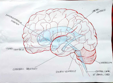
The brain contains four ventricles containing
cerebrospinal fluid (CSF).
They are right and left lateral ventricles, 3rd
ventricle and 4th ventricle.
Lateral ventricles lie on the 3rd
ventricle.
The 3rd ventricle is situated below the
lateral ventricles between the two parts of the thalamus. 3rd
ventricle communicates with the 4th ventricle by a canal called cerebral aqueduct.
The 4th ventricle is a diamond shaped
cavity situated below the 3rd ventricle between the cerebellum and
pons. The 4th ventricle is continuous below with the central canal of the spinal cord.
CEREBRO SPINAL FLUID (CSF)
CSF circulates constantly from the ventricles
through the sub arachnoid space (space between pia mater and arachnoid) around
the brain and the spinal cord.
CSF is a clear, slightly alkaline fluid, consisting
of water, mineral salts, glucose, HCo3-, oxygen etc.
CSF is secreted into each ventricle of the brain by choroid plexuses. Choroid plexuses are rich in blood
vessels and surrounded by ependymal cells in the
lining of ventricle walls.
From the 4th ventricle, CSF flows through
Foramen of luschka and foramen of magendie into the subarachnoid space and
completely surrounds the brain and the spinal cord.
FUNCTIONS
CSF protects the brain and the spinal cord from
mechanical shock by acting as a shock absorber.
CSF supplies glucose and oxygen to the brain.
SPINAL CORD
The spinal cord is an elongated part of the CNS.
It extends from the upper border of the 1st
cervical vertebra to the lower border of the 1st lumbar vertebra.
It is approximately 45cm long in adult males.
GREY MATTER OF SPINAL CORD
The arrangement of grey matter in the spinal cord
resembles the shape of the letter ‘H’.
The grey matter of the spinal cord has two posterior
(dorsal) horns and two anterior (ventral) horns.
Dorsal horns of grey matter of the spinal cord are composed of the cell bodies of sensory neurons, which carry information from the body to the brain.
Ventral horns of grey matter of the spinal cord are
composed of the cell bodies of the lower motor neurons, which carry back
information from the brain to the body.
WHITE MATTER OF SPINAL CORD
The white matter of the spinal cord consist of the
ascending tracts and descending tracts.
Ascending tracts are the axons of the sensory
neurons and descending tracts are the axons of the motor neurons.
REFLEX ARC
Spinal reflexes consist of three elements; sensory
neurons, interneurons in the spinal cord and lower motor neurons.
In the simplest reflex arc there is only one of each
of these neurons.
A reflex action is an involuntary and immediate
motor response to a sensory stimulus for protection.
Ex- knee jerk experiment


It's helpful
ReplyDelete