THE SENSORY ORGANS
PREPARED
BY MR. ABHIJIT DAS
THE EYE
Eyes are located in sockets of the skull called
orbits. The human eyeball is a spherical structure.
The eyeball wall is composed of three layers.

The external layer is
composed of dense connective tissue and is called sclera. The anterior portion
of sclera is called cornea.
The middle layer contains
many blood vessels and is called choroid. The choroid is bluish in colour. The
choroid layer becomes thick in the anterior part to form the ciliary body. The
ciliary body itself continues forward to form the iris which is the visible
coloured portion of the eye.
Eyes contain a transparent lens which is held by
ligaments attached to the ciliary body.
The hole located in the centre of the iris of the
eye that allows light to strike the retina is called pupil.
The inner layer is
the retina. Retina contains three layers of neural cells; photoreceptor layer,
bipolar layer and multipolar layer.
Photoreceptor layer consists of photoreceptor cells.
There are two types of photoreceptor cells; rods and
cones. These cells contain light sensitive
proteins called the photopigments.
The rods are responsible for twilight (scotopic)
vision and the cones are responsible for daylight (photopic) vision and colour
vision.
The rods contain a photopigment called the rhodopsin.
There are three types of cones which have their own
pigments that respond to red, green and blue lights.
The pigments respond to red light is erythropsin, the pigments respond to blue light is cyanopsin and the pigments respond to green light is chloropsin.
The sensation of different colours are produced by
various combinations of these cones and their pigments.
There is a region in the eyeball where the optic
nerves leave the eye and the retinal blood vessels enter the eye. Photoreceptor
cells are absent in that region and hence it is called the blind spot.
There is another region at the posterior pole of the
eye called macula lutea with a central pit called fovea. The fovea is the
location where only cones are densely packed and is called yellow spot. It is the point where the visual
resolution is the greatest.
The space between the cornea and the lens is the
aqueous chamber which contains a watery fluid called aqueous
humor.
The space between the lens and the retina is called
the vitreous chamber which contains a transparent gel called vitreous humor.
MECHANISM OF VISION
The light rays are focused on the retina through the
lens. These light rays generate action potential in the rods and cones.
The photopigments in the human eyes are composed of opsin (a protein) and retinal (an
aldehyde of vitamin A).
When light falls on photopigments , the retinal gets
separated from opsin and this process is known as bleaching
of pigments. This is how potential differences (action potentials) are
generated in the photoreceptor cells.
This produces a signal that generates action
potentials in the bipolar cells.
Then ultimately, the multipolar cells get
depolarized.
Now these action potentials are transmitted by the
optic nerves to the visual sensory area of the cerebrum, where the impulses are
analysed and the image formed on the retina is recognized based on earlier
memory.
THE EAR

The human ear can be divided into three major
sections; the outer ear, the middle ear, and the inner ear.
The outer ear
consists of the pinna and external auditory meatus (canal) and tympanic
membrane.
The pinna collects the vibration from the air which
produce sound. The external auditory meatus extends up to the tympanic membrane
(or the ear drum). There are very fine hair and ceruminous glands which produce
ear wax.
The tympanic membrane is composed of connective
tissues.
The middle ear contains
three ossicles (bones) called malleus, incus and stapes which are attached to
one another.
The malleus is attached to the tympanic membrane and
the stapes is attached to the oval window of the cochlea.
The ossicles present inside the inner ear increase
the efficiency of transmission of sound waves to the inner ear.
A tube called eustachian tube connects the middle
ear cavity with the pharynx. The tube helps in equalising the pressure on
either side of the tympanic membrane.
The inner ear is
called labyrinth which has two parts; the bony labyrinth and the membranous
labyrinth.
MECHANISM OF HEARING
The bony labyrinth of
the inner ear is a series of channels. Inside this bony labyrinth lies the membranous labyrinth, which is surrounded by a fluid
called perilymph and the membranous labyrinth is filled with a fluid called
endolymph.
The coiled part of the inner ear is called cochlea. Inside cochlea the upper membrane of the
membranous labyrinth is called the reissner’s membrane
and the lower membrane of the membranous labyrinth is called the basilar membrane.

A structure called the organ
of corti is located on the basilar membrane which contain hair cells that acts as auditory receptors. The basal
end of the hair cells are in close contact with nerve fibres.
Some projections called stereo
cilia are projected from the tip of each hair cells.
Another thin elastic membrane is present above the
hair cells called the tectorial membrane.

The external ear receives sound waves and transfers
the sound waves to the tympanic membrane.
The tympanic membrane vibrates in response to the
sound waves and these vibrations are transmitted through the ear bones
(malleus, incus and stapes) to the inner ear.
The vibrations are then passed to the fluid of the
cochlea, where they generate waves in the fluids.
The waves in the fluids induce a movement in the
basilar membrane. These movements of the basilar membrane bend the hair cells,
pressing their stereo cilia against the tectorial membrane.
As a result action potentials are generated in the
associated neurons present below the hair cells.
These action potentials are transmitted via auditory
nerves to the auditory sensory area of the cerebrum, where the impulses are
analysed and the sound is recognized.
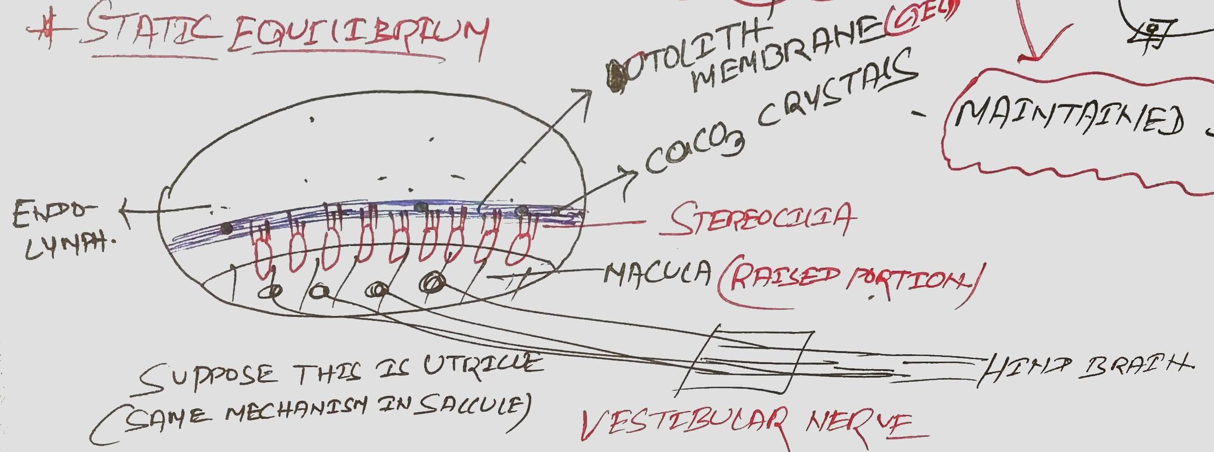
MECHANISM OF BALANCE
The inner ear also contains another structure called
vestibular apparatus composed of three
semicircular canals (bony) and the otolith organs (saccule and utricle).
Each semicircular canal lies in a different plane at
right angles to each other.
There are membranous canals which are suspended in
the perilymph of the semicircular canals.
The base of semicircular canal is swollen and is
called ampulla, which contains a raised
structure called crista ampullaris. Crista
ampullaris has hair cells. These hair cells are specific receptors of the
vestibular apparatus responsible for dynamic equilibrium
of the body.

The otolith organs also have raised structures
called macula. Macula has hair cells. These hair
cells act as receptors responsible for the static
equilibrium of the body.
So the semicircular canal and the otolith organs are
concerned with equilibrium of the body.
The arrangement of semicircular canals and otolith
organs allows perception not only of the position of the head in space but also
the direction and rate of any movement.
Any change of position of the head causes movement
in the endolymph containing the hair cells, which moves them and generates
action potentials in the sensory nerves present in the otolith organs and the
semicircular canals.
These action potentials are transmitted by the
vestibular nerve, which joins the cochlear nerve to form the vestibulocochlear
nerve.
The vestibular branch then passes to the cerebellum.
Thereafter, impulses are transmitted to the cerebrum and the skeletal muscles,
enabling perceptions of any adjustments needed to maintain balance or
equilibrium.
SENSE OF SMELL
The sense of smell or olfaction originates in the
nasal cavity.
MECHANISM OF SMELL
All odorous materials have volatile molecules, which
are carried into the nose by air.
When these molecules are dissolved in mucus, they
stimulate the olfactory nerves present in the mucus membrane of the roof of the
nasal cavity.
On each side of the nasal septum olfactory nerve
fibres pass to the olfactory bulb, where synapses occur.
From the olfactory bulb, bundles of nerve fibres
passes to the olfactory sensory area in the temporal lobe of the cerebrum,
where the impulses are interpreted and odour perceived.

THE SKIN
Skin is the largest sensory organ, and it plays a crucial role
in perception of the external environment. It is equipped with a variety of sensory receptors that allow to respond to
different stimuli. These sensory receptors are responsible for various types of
sensations, including touch, pressure, temperature,
pain, and even a sense of one's body position in space (proprioception).
The skin consists of three primary layers:
1.
Epidermis:
The epidermis is the outermost layer of the skin, serving as a protective
barrier between the body and the external environment. It is avascular (lacks blood vessels) and primarily
composed of stratified squamous epithelial cells.
Melanocytes within the epidermis produce
melanin, the pigment responsible for skin color.
2.
Dermis:
The dermis is the layer beneath the epidermis and is primarily composed of connective tissue. It provides structural strength
and elasticity to the skin. The dermis contains a network of collagen and
elastin fibers, which give the skin its flexibility and resilience. This layer
contains various supporting structures, such as hair
follicles, sweat glands, sebaceous (oil) glands, blood vessels.
3.
Subcutaneous Tissue (Hypodermis):
The subcutaneous tissue, also known as the hypodermis
or superficial fascia, is located beneath the dermis. It consists mainly
of fat cells (adipocytes) and connective tissue. The hypodermis provides
insulation and padding, helping to regulate body temperature and protect
underlying structures.
INJECTIONS ADMINISTERED
INTO THE SKIN
1.
Intradermal Injections:
These injections are administered into the dermis of
the skin. Intradermal injections are typically given at an angle (5-15 degrees) and are often used for
allergy testing and some vaccines, such as the tuberculosis (TB) skin test.
2.
Subcutaneous Injections:
Subcutaneous injections are delivered just beneath the skin into the subcutaneous tissue. The needle is
typically inserted at a 45-degree angle to
the skin. Subcutaneous injections are commonly used for medications that need
to be absorbed slowly, like insulin or some vaccines.
3.
Intramuscular Injections:
Intramuscular injections are administered into the muscle
tissue beneath the skin. In adults, a 90-degree
angle (perpendicular to the skin) is typically used. Intramuscular
injections are often used for a wide range of medications, including vaccines,
antibiotics, and certain hormones.