HUMAN REPRODUCTIVE SYSTEM
PREPARED BY MR. ABHIJIT DAS
MALE REPRODUCTIVE SYSTEM
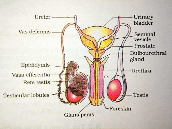
The male reproductive system has three main parts: the
primary sex organ (testes), accessory ducts and glands, and external
genitalia (penis).
TESTES
The testes are located in a pouch-like
structure called the scrotum. Each testis has about 250 compartments
called testicular lobules. Each lobule contains 1-3 seminiferous
tubules where sperm are produced. These tubules have two main types of
cells:
- Spermatogonia
– undergo division to form sperm.
- Sertoli
cells – provide support for sperm development.
The testes also contain interstitial cells or Leydig
cells, which produce testosterone.
ACCESSORY DUCTS AND GLANDS
The male sex accessory ducts include tubuli recti,
rete testis, vasa efferentia, epididymis, and vas
deferens. The vas deferens loops over the bladder, joins with a duct from
the seminal vesicle, and opens into the urethra as the ejaculatory
duct. The prostate gland and bulbourethral gland also release
their secretions into the ejaculatory duct.
Semen is a mix of sperm
and secretions from the seminal vesicle, prostate gland, and bulbourethral
gland.
EXTERNAL GENITALIA
The penis is the male external genitalia. It
contains two types of erectile tissues: corpora cavernosa and corpus
spongiosum. The visible tip is called the glans penis, which has Tyson's
glands for lubrication.
FUNCTIONS OF TESTOSTERONE
Testosterone
has several key functions:
1.
Stimulates sperm production.
2.
Promotes male secondary sexual
characteristics like beard growth, deep voice, and broad chest.
3.
Has an anabolic effect on bones and
muscles, promoting their growth.
FEMALE REPRODUCTIVE SYSTEM
The female reproductive system includes a pair
of ovaries, a pair of oviducts (fallopian tubes), the uterus,
cervix, vagina, external genitalia, and a pair of mammary
glands.
The ovaries are the primary sex organs in
females. They produce female gametes (eggs) and release female
hormones. Each ovary
is about 2-4 cm in length.
Each fallopian tube is 10-12 cm long and has three parts: the infundibulum,
ampulla, and isthmus. The edge of the infundibulum has
finger-like projections called fimbriae, which help in collecting the ovum
from the ovary.
The uterus
(womb) has three layers: perimetrium (outer), myometrium
(muscular middle), and endometrium (inner lining).
The cervical
canal and vagina together form the birth canal.
The external
genitalia consists of the mons pubis, labia majora, labia
minora, hymen, and clitoris.
MAMMARY
GLAND:
The mammary
glands (breasts) are paired structures containing fat and 15-20
lobules. Each lobule has clusters of cells called alveoli, which
secrete milk.
The mammary ducts gather milk from lobules. These ducts join into a mammary ampulla, which connects to the lactiferous duct at the nipple, where milk is released.
SPERMATOGENESIS
1.
Spermatogonia
(stem cells) are located inside the seminiferous tubules.
2.
They
undergo mitosis, forming two cells:
o One remains as spermatogonia
to maintain the reserve.
o The other becomes a primary
spermatocyte.
3.
Primary spermatocytes undergo meiosis I, forming two secondary spermatocytes.
4.
Secondary spermatocytes undergo meiosis II, producing spermatids
(haploid).
5.
Spermatids
undergo maturation (spermiogenesis) to form spermatozoa (sperm).
OOGENESIS
1.
Begins
in the ovary during fetal development.
2.
Oogonia (stem
cells) divide by mitosis to form primary oocytes.
3.
Primary
oocytes enter meiosis I but pause at prophase I until puberty.
4.
At
puberty, one primary oocyte resumes meiosis I during each cycle, forming:
o A large secondary oocyte
o A small polar body
(discarded).
5.
The
secondary oocyte begins meiosis II but pauses at metaphase II
until fertilization.
6.
If
fertilization occurs, meiosis II completes, forming:
o A mature ovum
o Another polar body.
7.
If
no fertilization, the secondary oocyte degenerates, and the cycle restarts.
MENSTRUAL CYCLE
1.
Duration:
About 28 days (varies from 21-35 days).
2.
Phases:
o Menstrual Phase (1-5 days): Shedding of the endometrial
lining causes bleeding.
o Follicular Phase (6-13 days): Follicles in the ovary grow;
estrogen levels increase; endometrium regenerates.
o Ovulation (Day 14): A mature egg is released from the ovary
due to a spike in LH (Luteinizing Hormone).
o Luteal Phase (15-28 days): The corpus luteum forms and
secretes progesterone to maintain the endometrium.
3.
If fertilization occurs: The embryo implants in the endometrium.
4.
If no fertilization: The corpus luteum, along with the endometrium,
degenerates, hormone levels drop, and the cycle restarts.
PREGNANCY
Pregnancy is the time
during which a baby develops inside the mother’s womb, lasting about 40 weeks.
STAGES OF PREGNANCY:
1.
First Trimester (0–12 weeks):
o Fertilization and implantation
occur.
o The
embryo develops major organs and systems (heart,
brain, and spinal
cord).
o By
the end of this stage, the embryo becomes a fetus.
o Common
maternal symptoms: nausea, fatigue, and hormonal
changes.
2.
Second Trimester (13–26 weeks):
o The
fetus grows significantly; facial features and
limbs develop further.
o Movements (quickening) are felt by the mother around
18–20 weeks.
o Development
of organs like lungs and bones progresses.
o Maternal
symptoms: reduced nausea and visible baby bump.
3.
Third Trimester (27–40 weeks):
o The
fetus gains weight.
o Common
maternal symptoms: back pain, frequent urination, and Braxton
Hicks contractions (painless uterine contractions).
PARTURITION
1.
It is the process
of childbirth.
2.
Divided into three stages:
o Dilation:
Opening of the cervix.
o Expulsion:
Delivery of the baby.
o Placental
Stage: Delivery of the placenta.
ROLE OF OXYTOCIN IN PARTURITION
1.
Stimulates strong
uterine contractions to facilitate the process of childbirth.
2.
Reduces postpartum bleeding by
contracting the uterus.


